Oncology Decision Support Software
Special laboratory of IT specialists and medics supported by Sechenov university, one of the most important medical universities in Russia, have developed an AI that allows to increase the efficiency of treatment of colorectal cancer. The neural network is trained to map areas on digital images of histological samples and identify tumors and metastases 88% faster than doctors could do it before technology implementation.
I was part of team and was responsible for end-to-end development of MVP version featuring general functionality for medical specialists.
I was part of team and was responsible for end-to-end development of MVP version featuring general functionality for medical specialists.
Doctors would like to have shared access to process scans together
and train less experienced colleagues
and train less experienced colleagues
Shared studies
There is a hierarchy of specialists inside the laboratory, each level needs its own functionality inside the software
Suitable for all
To create an MVP with the functionality of an image viewer and data tables with management on behalf of a Pathomorphologist user to speed up the process of making medical decisions and choosing treatment tactics, reducing routine tasks for medical workers. Medical professionals operate in high-pressure environments where precision and efficiency are critical, making it essential to design a tool that seamlessly integrates into their routine.
Challenge
Current processing and marking of the scan takes a lot of time and slows down the processes
Fast process
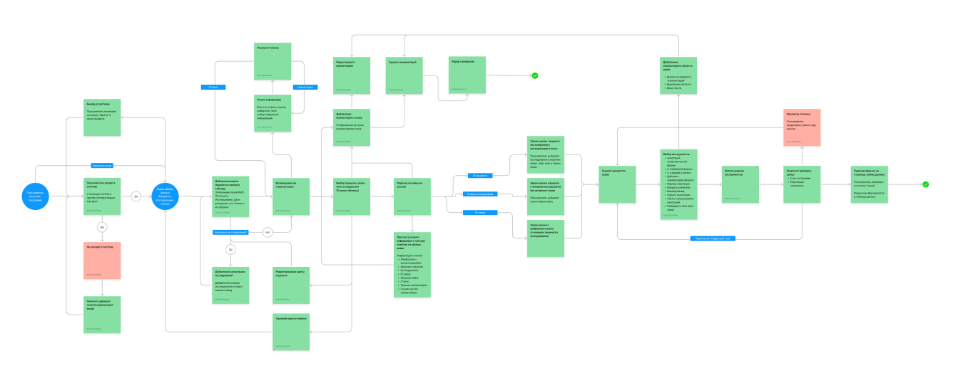
Pathomorphologist User Flow
They would like to work with a simpler interface so that they don't have to study the instructions for the program
Simple interface
After understanding the challenges and needs of doctors, I created a User Flow for the pathologist – the primary user who handles clinical data and accesses a database of histological scans. Once the basic structure of the software was outlined, including the functionality and tools for viewing scans, I designed the key screens as wireframes.
User Flow
Research
At the beginning, me and product team conducted 2 interviews with the Head of laboratory and doctors and found out about doctors needs:
Benchmarking
I analyzed 5 AI-driven healthcare applications to understand how they handle user workflows, AI integration, specific features like commenting and team work. A common best practice across top apps was to use toolbar and sidebars for the viewer page allowing user close interaction and switching between scans. Important point was to show overview of AI results and let specialists see all active scans and their preview. Based on the findings, I prioritized creating an intuitive and responsive visualization of scans data and incorporate a collaborative workspace features
Wireframes
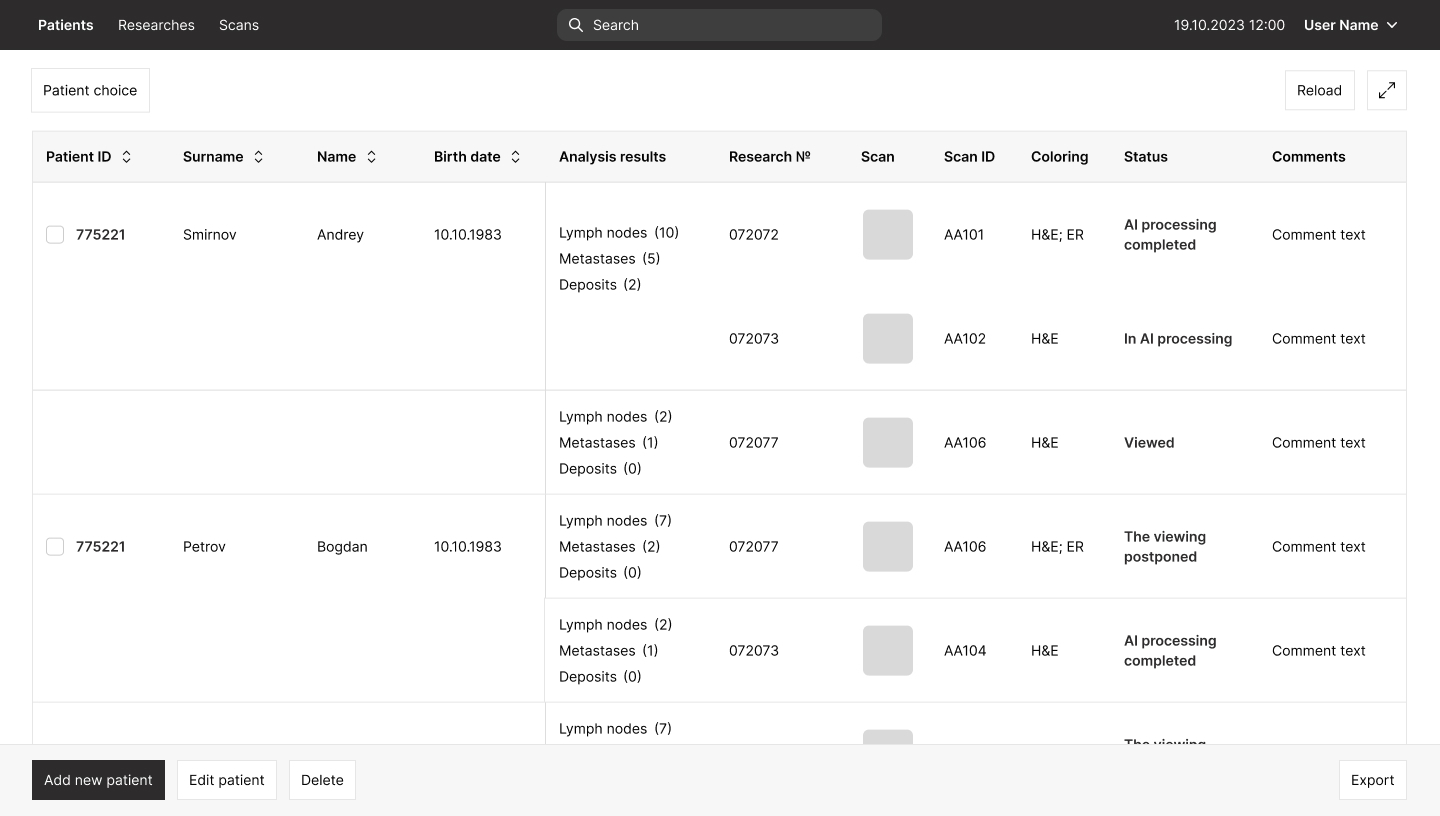
First layouts
On the first versions of the layouts, it was suggested to add a filter by clients, make a preview of comments in the table. The final versions have changed after technical reviews and UX tests
First versions
After forming the overall structure, we started looking for a core and cutting functionality for the first iteration of development. This is how the first version of MVP and the core of the product were assembled, which was ready for testing on real users.
Tech-review
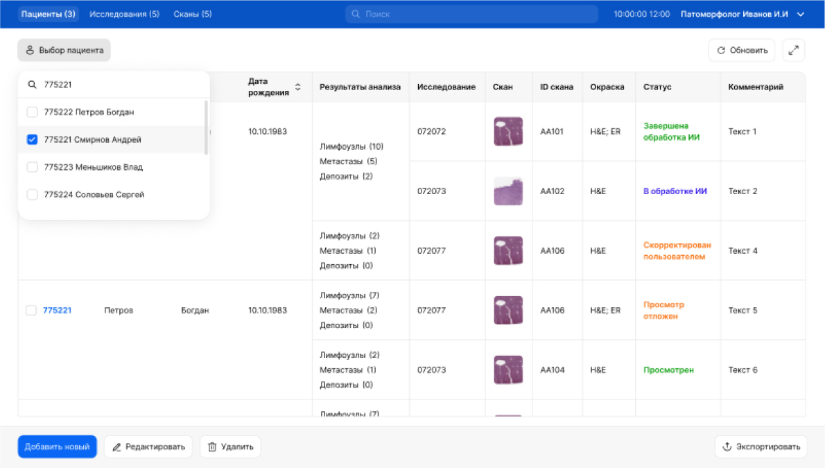
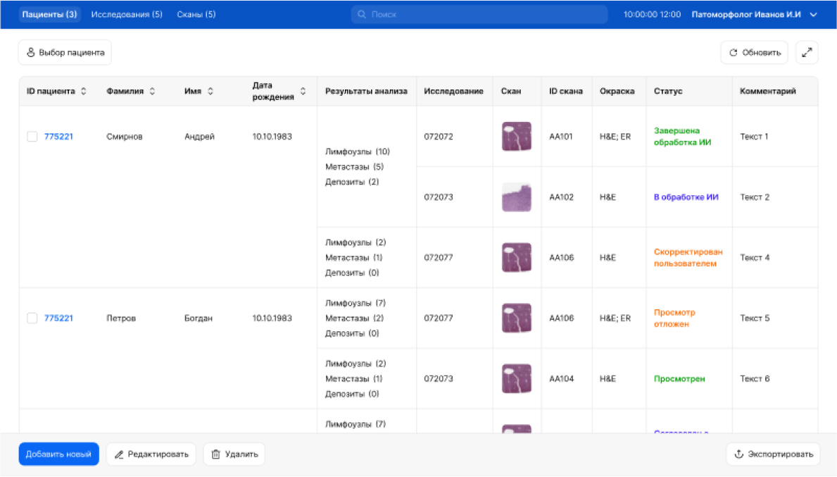
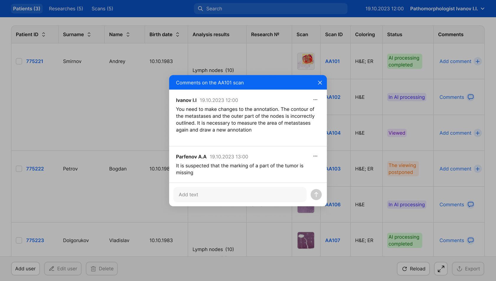
The main table is interactive field and consists everything doctors need including scan preview that can be zoomed on click and the possibilty to add comments and see already existing ones.
#1 One table for everything
Comments history - Table scenario
Users journey begins on the front page of patiens. I decided to present this page in table form because doctors found this structure the most easy to understand like they always did using other programs.

Solutions
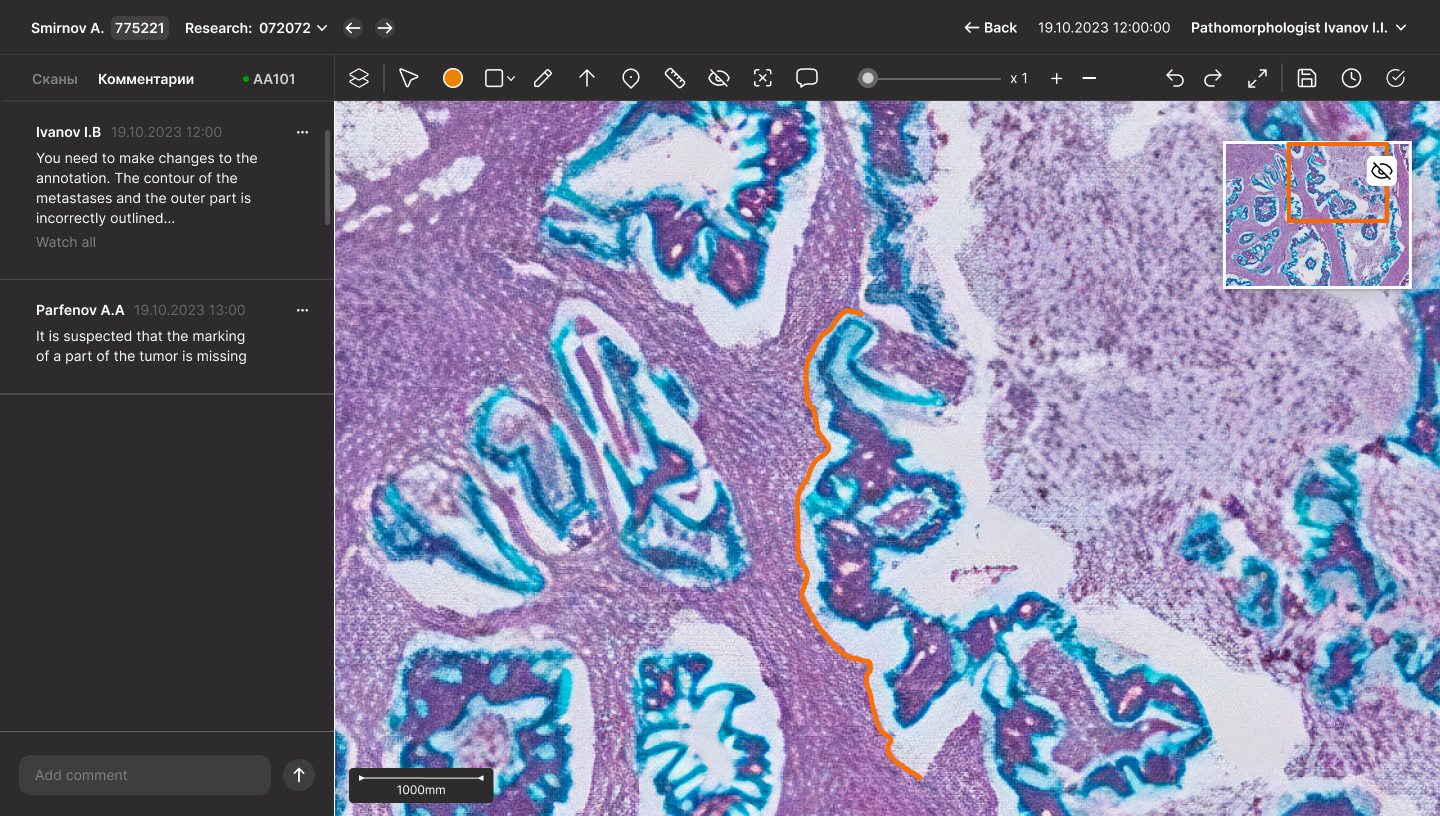
Scan viewer comment tab
#2 Flexible workspace
The comment tool allows users to add remarks directly on specific points of the scan or on the entire scan as a whole. This ensures thorough attention to detail and enhances collaboration during teamwork.
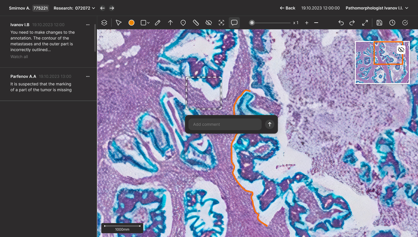
Add comment
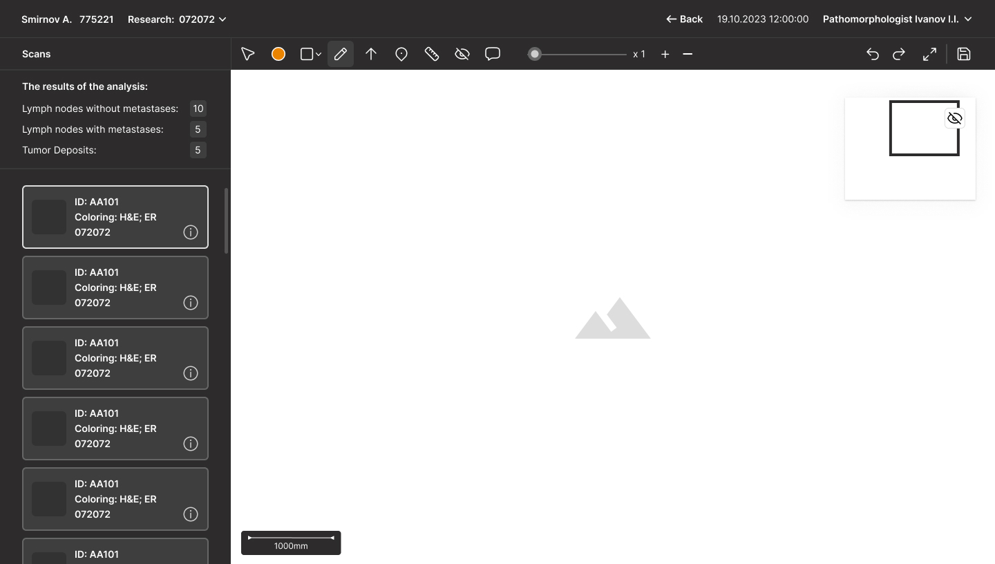
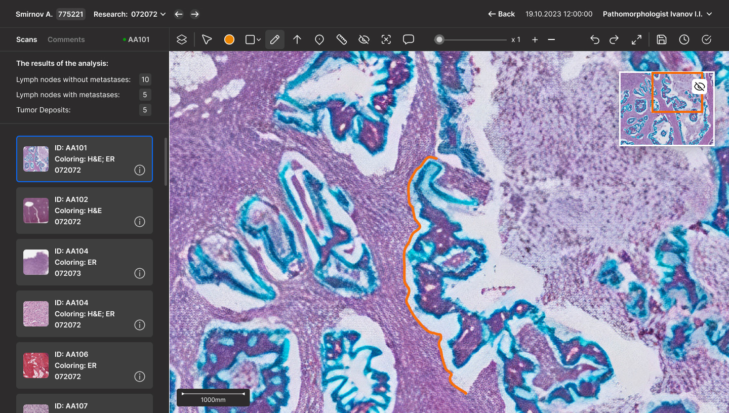
The scan viewer is a dedicated workspace where doctors can review AI analysis results, add their own marks and measurements, and collaborate with others through comments. Designed for efficiency, it eliminates unnecessary steps to switch between tabs and tools. Its structure, inspired by best practices, ensures users can easily locate and access the tools they need.
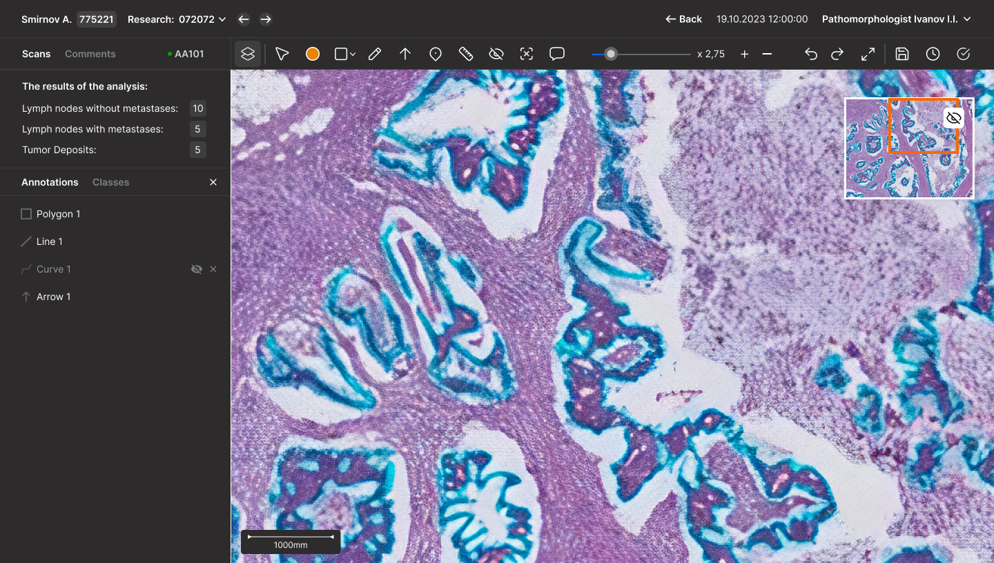
Layers tab
#3 Need for more editing
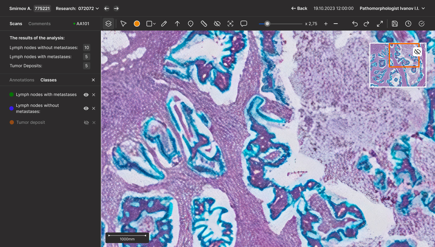
Classes tab
This software already has found its users in the Laboratory of Sechenov University and gets tested, it was presented on multiple medical conferences featuring AI technologies and has been highly appreciated
The scan workspace is a graphical interface that closely resembles those used by designers. To enhance usability, I designed a layers functionality accessible via the toolbar. This feature allows users to activate layers (annotations) and manage classes, which are lists of marks with histological definitions. These can be hidden or deleted as needed, providing flexibility and ease of use.